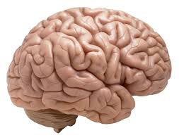
Human brain, command center for the nervous system, still holds the mystery for its folds in the history of medical sciences.While many researchers believe that the folds of the brain exist to increase the surface area and fit into the skull space, there is no evidence to prove this.The researchers conduct many live experiments on the brain to know the reason behind those folds.One such experiment was done using 3D printing technique that mimicked the growth and development of the brain.
The researchers’ experiments with 3D printed brain stimulation proved that folds are caused by mechanical compression forces in response to the brain’s rate of growth.As the age of a human body increases, the number of folds in the brain also increases; this process is called as gyrification.The researchers proved that the folded structure is simply a physical growth process and not a matter of biology.
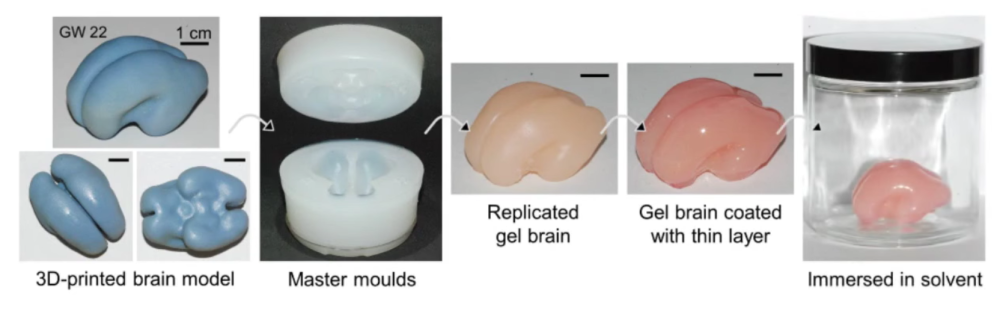
The stimulation of brain development modal was based on MRI data that used several layers of soft gel materials. The layers of gel were designed to expand and swell at different rates when placed in a specialized liquid solution, The process was designed to simulate the growth and development of the brain’s gyri and sulci, the areas of folded tissue on the surface of the brain. The 3D printed model of the brain accurately develops folds like the real human brain.
“Starting with the same initial geometry, we also build numerical simulations of the brain modelled as a soft tissue with a growing cortex and show that this also produces the characteristic patterns of convolutions over a realistic developmental course. All together our results show that although many molecular determinants control the tangential expansion of the cortex, the size, shape, placement and orientation of the folds arise through iterations and variations of an elementary mechanical instability modulated by early foetal brain geometry,” wrote the study authors Tuomas Tallinn, Jun Young Chung, François Rousseau, Nadine Girard, Julien Lefèvre and L. Mahadevan.
This research helps for early detection and treatment of neurological disorders, malfunctioning of brain, cortex thickness disorders, etc. This model is limited only to predicting the behavior of simple, basic brain structures at the very onset of the folding process.
“Their simulations explain why folding always begins in weakly curved regions and why gyri and sulci align perpendicular to the direction of maximum compressive stress. Experiments with swelling brain models provide the essential missing link between modelling, experiment and simulation. However, a few limitations remain: the model is beautiful and simple, but it is limited to the initial folding of idealised structures; the experiment is useful for exploring instabilities beyond the onset of folding, but it is limited to moderate changes in volume,” explained the Departments of Mechanical Engineering and Bioengineering at Stanford University in California’s Ellen Kuhl in an accompanying paper that she authored, also published by Nature Physics.
You can see a video overview of the experiment here:
https://youtu.be/gbSGIG9SCf8
The experiment can lead to new medical advancements in treating neurological disorders . Specifically, being able to link the rate of brain growth to neurological development could help scientists trace individual brain functions back to the folding of the brain surface. Because the process is mechanical, not biological, it could accurately determine how and when something goes wrong. This could lead to the ability to identify surface markers that may lead to the early diagnosis of autism, schizophrenia or Alzheimer’s disease. Early detection could also lead to the development of more effective treatment options and the cultivation of new preventative measures.