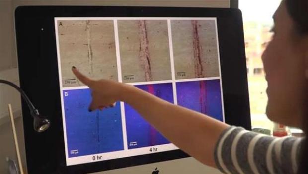
X-rays have been around since 1895. They are the best ways to determine the condition of the bones. Recently the chemical researchers at Trinity College and the Royal College of Surgeons in Dublin have found a new 3D scanning method. As per this scanning tool, gold will be pumped into the blood. When a bone is injured or cracked, it introduces some amounts of calcium into the blood. A luminescent material can be attached to the gold particles which get drawn towards the calcium deposits.
A scan will thereafter, be carried out to determine the level of crack and injury in the bones. Using this technique, the slightest of problems in the bone can be detected. Although in this scanning method, a foreign object is introduced into the blood, it is safe.
Gold is extensively used in healthcare services as it is stable and non-reactive. X-rays cause cancer and this method is a safer alternative. It does not contain any radiation rays. Apart from highlighting injuries in the bones, this technique could well be used to identify the onset of bone diseases. Preventive measures can be taken thereafter to prevent bone issues.
Clive Lee is a professor of Anatomy at the Royal College of Surgeons. He has supervised the team in the research of the 3D scanning process. He says that the biggest advantage of this technique will be identifying the problem, rather than treating the cracks. Micro cracks develop in the bones, and with increased activity and load, the bones weaken. Eventually, this leads to injury. Identification in the early stages will be beneficial for athletes and elderly people. With no harmful elements like x-rays, this technique might be safer. It is yet to ascertain the cost of the scanning system.
Source: 3ders.org